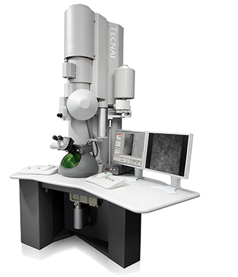
Transmission Electron Microscopy (TEM)
Transmission electron microscopy is a microscopic technique in which an electron beam is transmitted through a thin specimen. Interaction of energetic electrons with the specimen in the vacuum chamber produces a high-resolution, black and white image on an imaging device, such as a fluorescent screen, on a layer of photographic film, or to be detected by a sensor such as a CCD camera. The TEM has the added advantage of greater resolution - in hi-res even as small as a single column of atoms, owing to the small de Broglie wavelength of electrons.` Alternate modes of TEM (STEM, HAADF, EDS) allow to observe modulations in chemical identity, crystal orientation, electronic structure and sample induced electron phase shift as well as the regular absorption based imaging.
In our lab, nanoparticles (mineral oxides and engineered particles) are characterized with various modes of TEM to obtain particle size distributions, shape information and elemental compositions. Through collaborations, we have access to FEI Tecnai G(2) F30 S-Twin 300 kV transmission electron microscope at the Center for Integrated Nanotechnologies (CINT), Los Alamos. This produces 0.2 nm point resolution in TEM and 0.19 nm in high angle annular dark field (HAADF) Scanning TEM and is equipped with an EDAX ECON x-ray detector, Gatan GIF Tridiem electron energy loss spectrometer, Gatan Ultrascan 1000 CCD camera, Gatan bright field, Gatan annular dark field, and Fischione Instruments HAADF STEM detectors.

ADMINISTRATIVE ASSISTANT
GAYAN R. RUBASINGHEGE
Associate Professor of Chemistry
New Mexico Institute of Mining and Technology
Department of Chemistry
801 Leroy Place
Socorro, NM 87801
Bethany Jessen
New Mexico Institute of Mining and Technology
Department of Chemistry
801 Leroy Place
Socorro, NM 87801
Phone: 575-835-5129
Fax: 575-835-5215
Phone: 575-835-5263
Fax: 575-835-5364
Copyright © 2018 The Environmental Chemistry Research Research Group. All rights reserved.